Volume 05
Issue 02
May 2017
Inside This Issue
Opinion/Editorial, 2
Technology Corner, 3,4
Tips from the Experts, 5,6
Humanitarian News, 7
Educaon and Training, 8,9
BOR News, 10
Research, 11
Bronchoscopy Around the World, 12
WABIP Academy Webcasts, 13
Links, 13
Upcoming Events, 14

a lack of PCP commitment to LCS.
5
Planning also requires the support
of local leadership and a business
model that includes funding for a
nurse navigator and database devel-
opment or soware plaorm to
manage and track screened pa-
ents, nodules detected, and allows
for data reporng to an accredited
registry. The implementaon phase
should emphasize how to ensure
that screening is only performed in
appropriate individuals, how to per-
form shared decision making and
incorporate tobacco cessaon, the
process for following up abnormal
ndings, and adherence to repeat
imaging. Lastly, maintaining the pro-
gram should involve reviewing quali-
ty metrics and registry data to en-
sure it is operang as intended.
In conclusion, implemenng LCS has
many moving parts with challenges
that may vary based on locally avail-
able resources and enthusiasm for
screening, but it absolutely can be
done. To develop and implement a
program that is eecve and safe
involves buy in from many dierent
disciplines and services. A carefully
planned approach with a focus on
the essenal components for LCS
will do much to ensure a successful
program start and uptake. Finally,
connuing review of system and
paent level outcomes is important
for quality assessment and future
adaptaons of the program.
References
1. Aberle DR et al. N Engl J Med.
2011;365(5):395-409.
2. Bach PB et al. JAMA. 2012;307
(22):2418-29.
3. Mazzone P et al. Chest. 2015;147
(2):295-303.
4. Wiener RS et al. Am J Respir Crit Care
Med. 2015;192(7):881-91.
5. Gesthalter YB et al. Chest. 2017
Nichole T. Tanner,
MD, MSCR
Associate Professor of
Medicine
Co-Director, Lung Cancer
Screening Program
Medical University of
South Carolina
Core invesgator and Lung Cancer Screening
Director
Health Equity and Rural Outreach Innovaon
Center (HEROIC)
Ralph H. Johnson Veterans Aairs Hospital
Six years ago the landmark Naonal
Lung Screening Trial (NLST) was pub-
lished demonstrang a mortality
benet to screening asymptomac
individuals at high risk based on age
and smoking history with annual low
-dose computed tomography
(LDCT).
1
The number needed to
screen to prevent one death from
lung cancer was 320; a number simi-
lar to that for mammography in
women 60 and older. The NLST also
demonstrated a high number of false
posive results with LDCT screening
with approximately 1 in 4 paents
having a screen detected nodule. The
vast majority of these nodules (96%)
were not malignant in nature and
the potenal risk of downstream
invasive tesng for benign disease
along with paent anxiety gave many
pause to recommend widespread
implementaon of lung cancer
screening.
2
It wasn’t unl 2013 that the United
States Preventave Services Task
Force gave a lung cancer screening
(LCS) with LDCT a grade B recom-
mendaon for high risk individuals.
Following this recommendaon,
broad uptake of LCS did not occur as
many sll had concerns about the
best way to implement LCS and in-
surers were largely not providing
coverage. In March 2016, close to 5
years aer the publicaon of the
NLST, the Centers for Medicare and
Medicaid Services (CMS) approved
coverage for lung cancer screening
for its eligible beneciaries, however
nong the potenal risks, a paent
shared-decision making visit was
mandated prior to LDCT; the rst for
any cancer screening test.
Implemenng lung cancer screening
has become much more than adver-
sing and a scanner; professional
sociees cauon that LCS should be
conducted in a muldisciplinary and
comprehensive program that incor-
porates experse in pulmonary nod-
ule management as well as tobacco
treatment services. In a joint policy
statement, the American College of
Chest Physicians and the American
Thoracic society recommend nine
programmac components to ensure
that LCS is conducted eecvely, with
quality, and safety.
3
These compo-
nents include standardized protocols
for performing LDCT, reporng re-
sults, and pulmonary nodule evalua-
on. Paent eligibility, frequency and
duraon for LCS comprise as well as
paent and provider educaon are
addional components.
While these essenal components
provide an ideal framework for im-
plementaon, the real-world logiscs
of starng a LCS program can be com-
plicated. The ACCP and ATS outline
strategies for the successful imple-
mentaon LDCT screening programs
into clinical pracce in a separate
policy statement.
4
These praccal
approaches are categorized into
three phases: planning, implementa-
on, and maintenance of LCS. The
planning phase should be guided by a
muldisciplinary steering commiee
that includes engagement and educa-
on of primary care providers. Evalu-
aons of early-adopng LCS pro-
grams at three unique centers sug-
gests that failure to do so resulted in
Guest Opinion/Editorial
WABIP Newsletter
M A Y 2 0 1 7 V O L U M E 5 , I S S U E 2
EXECUTIVE BOARD
Zsolt Papai MD
Székesfehérvár, Hun-
gary
Chair
Silvia Quadrelli MD
Buenos Aires, Argen-
na
Vice-chair
Hideo Saka MD
Nagoya, Japan
Secretary General
Hojoong Kim MD
Seoul, Korea
Treasurer
Eric Edell MD
Rochester MN, USA
President WCBIP 2018
Quangfa Wang MD
Beijing, China
President WCBIP 2020
Henri Colt MD
Laguna Beach, CA
Immediate Past-chair
STAFF
Michael Mendoza
General Manager
Judy McConnell
Administrator
Kazuhiro Yasufuku
Newsleer Editor-in-
chief
P A G E 2
How to set up a lung cancer screening program: more than a glossy brochure and a CT scanner

Technology Corner
Endobronchial blockers for Lung Isolaon in Massive Hemoptysis
Introducon: Balloon-based endobronchial blockers were developed for lung isolaon for thoracic surgical procedures as an alter-
nave to double lumen intubaon. They have been used, however, for lung isolaon in the seng of massive airway bleeding.
There are a variety of endobronchial blockers available on the market, which vary in regards to design and instrucons for use. The
purpose of this essay is to describe the basic specicaons and techniques for using two such devices as they may be applicable to
clinicians encountering massive airway bleeding.
Background: Most paents with massive hemoptysis die due to asphyxiaon. Approximately 150 to 200 milliliters (ml) of blood
could interfere with gas exchange and cause respiratory failure and death. This is because the amount of blood needed to ll the
anatomical dead space in most paents is only about 150 ml. Massive hemoptysis has been dened in dierent series as > 200 to >
600 mL of blood per 24 hours, but due to the rather small quanty of blood needed to ll the anatomic dead space, death will typi-
cally occur prior to exsanguinaon. Bleeding rate of ≥ 1,000 ml within a 24-hour period, aspiraon of blood in the contralateral
lung, massive bleeding requiring single-lung venlaon and lung cancer as underlying eologies have all been associated with high-
er mortality. Morbidity and mortality are reportedly less when tuberculosis, bronchis or bronchiectasis were responsible for the
massive hemoptysis. Some series report higher mortality rates in paents who experienced recurrent bleeding following bronchial
artery embolizaon (BAE) for massive hemoptysis.
The rst priority in the management of massive hemoptysis is to maintain a paent airway. While seng up the equipment for
endotracheal intubaon and lung isolaon, the paent should be placed in the lateral safety posion (lateral decubitus posion)
with the bleeding side down so blood does not also ll the unaected lung. Bronchoscopists should be familiar with the use of en-
dobronchial blocker placement as a means to isolate the bleeding lung, control massive hemoptysis and spare airways for gas ex-
change. In fact, transporng a paent to intervenonal radiology or intensive care unit without a secured airway (and isolated
bleeding lung) in cases of massive hemoptysis is considered unsafe in the event of airway occlusion from large blood clots in route.
Commonly used endobronchial blockers mainly vary in steering technique, balloon size, locking mechanism and method of place-
ment. The two blockers described herein need visual guidance for proper placement in the desired airway. A locking system is
available to secure the blocker in the desired locaon and reduce the risk for migraon. There are no published surveys, however,
assessing the operators’ comfort level or user-friendly features for the various available systems. The Arndt endobronchial blocker
(Cook Medical) require the use of a pediatric bronchoscope when the blocker is placed though a regular 7.0-8.5 endotracheal
tubes. An alternave technique can be used, in which the blocker can be inserted in the airway alongside the endotracheal tube,
even by using a regular diagnosc adult bronchoscope (Figure1). The VivaSight –EB (Ambu A/S) can be used without the broncho-
scope when inserted through a dedicated endotracheal tube with a built-in camera (VivaSight –SL, Ambu A/S) (Figure 2). To date,
there are no comparison studies between the blockers designed by dierent manufacturers. Familiarity, availability, the feasibility
of using a bronchoscope in emergent situaons and costs impact operators’ selecon of a parcular endobronchial blocker.
Clinical applicaons: The Arndt endobronchial blocker (Cook Medical) and the VivaSight-EB (Ambu A/S) while approved for clinical
use for lung isolaon, have not yet been systemacally studied for massive hemoptysis. There are several issues that require aen-
on when using these devices for lung isolaon in massive hemoptysis:
1. Massive bleeding from the le lung: selecve intubaon of the right main bronchus (RMB) should be performed emergently.
However, because of the short length of the RMB (1.5-2 cm), it is very likely that the takeo of the right upper lobe bronchus
would be occluded if the ETT is properly posioned in the RMB. Venlaon to the right lower lobe (RLL) and right middle lobe
(RML) may not be tolerated, and thus, alternaves have been proposed; a double lumen endotracheal tube could be consid-
ered, but it may not be feasible to place these tubes in emergent situaons for hemoptysis. Thus, an endobronchial blocker
could be placed in the le main bronchus, while keeping the ETT in the trachea (Figure 3). This way, the enre right lung is
being venlated, while prevenng spillage of blood from the le lung.
2. Massive bleeding from the right lung: in this scenario, the le lung should be selecvely intubated; this is feasible as the le
main stem bronchus is typically 5 cm in length and a single lumen ETT can be placed in posioned in the LMB. If the bleeding is
from the RML or RLL, however, the ETT can be secured in mid trachea and the blocker posioned in the Bronchus intermedius
(Figure 1), allowing venlaon not only of the le lung but also of the RUL.
W A B I P N E W S L E T T E R
P A G E 3
3. Paent safety during and blocker inseron:
A. The balloon should never be overinated; in fact, the balloon should be deated for a few minutes three mes a day in order to
preserve mucosal viability and to check for bleeding recurrence.
B. If venlaon becomes dicult during endobronchial blockade, the balloon should be deated and the its posion inspected as
migraon is possible
C. Higher PEEP and low dal volume may occur during placement due to the presence of scope and blocker in the ETT
Conclusions: The available endobronchial blockers have dierent design, inseron technique and maneuverability. The lack of published
literature makes a fair comparison between dierent blockers in the same paent populaon impossible. We believe that by appropriately
using the endobronchial blockers in the seng of massive hemoptysis, praconers can safely isolate the bleeding lung and potenally
stabilize paents unl denive treatment is oered.
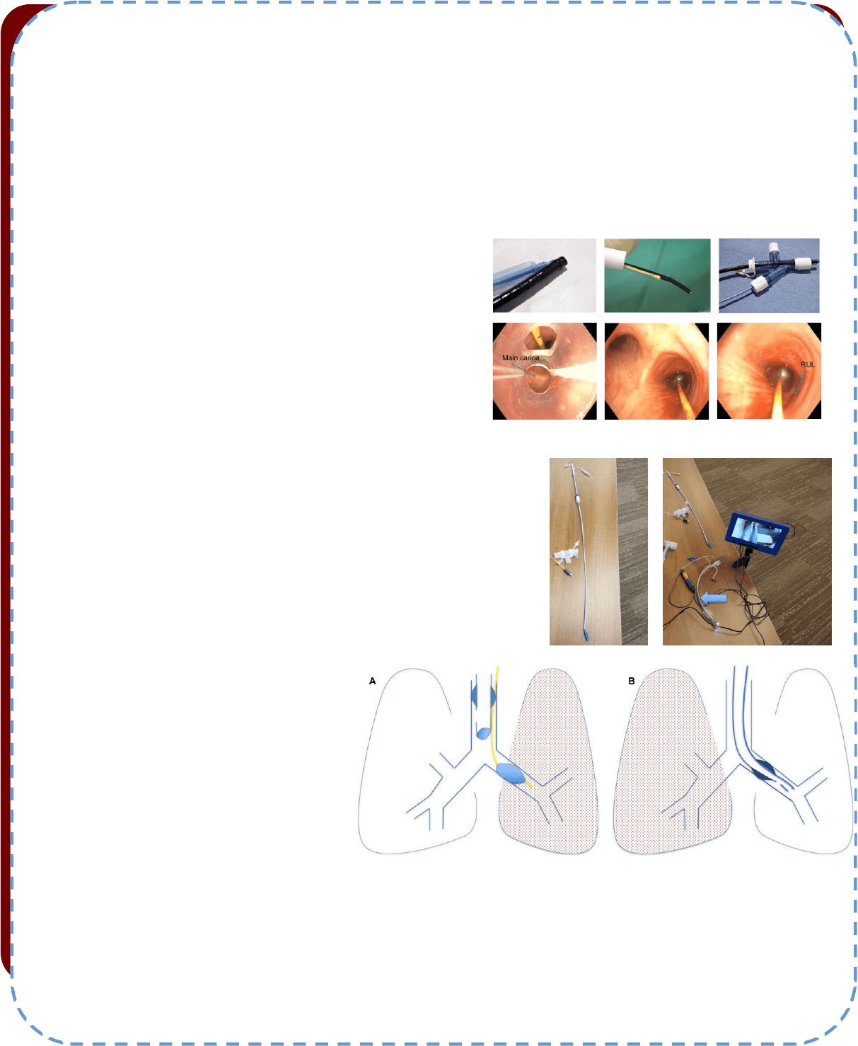
Figure 1
Arndt Endobronchial blocker. Top le: The Arndt endobronchial blocker uses a
guide loop assembly that ts through the lumen of the blocker and exits from the
blocker’s distal end to form a small, adjustable loop. Top center: The bronchoscope
is placed through the diaphragm of the bronchoscopy port of the Arndt Mulport
Airway Adapter; the bronchoscope is advanced through the guide loop. Top right:
Once coupled through the Arndt Mulport Airway Adapter, the bronchoscope and
the blocker are placedon the endotracheal tube and the paent venlated with
100% oxygen. The guide loop should be adjusted to loosely approximate the diame-
ter of the bronchoscope. Boom le: The blocker is inserted in the airway alongside
the endotracheal tube. Boom Center: The blocker can be placed in the bronchus
intermedius (BI) in cases of bleeding form the RML or RLL. Boom right: When
placed in the BI, the RUL bronchus patency is maintained. Photos courtesy of Eric Edell, Mayo Clinic and Sepmiu Murgu, University of Chicago.
Figure 2
Le panel: VivaSight-EB is an endobronchial blocker designated for lung isolaon. It consists of a
sterile, single-use, “steerable” balloon-pped catheter that is visually guided to a selected airway.
The angled distal p of VivaSight-EB can be adjusted to facilitate placement in the desired bronchi.
When used in conjuncon with the VivaSight-SL connuous monitoring throughout the procedure
ensures that dislocaon can be easily detected and corrected. Right panel: For visual guidance
during posioning the blocker can be used in combinaon with the bronchoscope or the VivaSight-
SL single lumen tube (arrow) with integrated camera.
Figure 3
Managing strategies for massive hemoptysis. A. In case of le
lung bleeding, the ETT can be secured in the trachea and the
endobronchial blocker placed either through the ETT or along
its side and posioned in the le main bronchus. The right
upper lobe can be closed o with a right mainstem bronchus
intubaon. B. In cases of right lung bleeding, the le main
bronchus can be intubated over the bronchoscope. This is
possible as the LMB is ~5 cm in length.
References
1. Sakr L et al. Respiraon 2010; 80: 38-58
2. Shigemura N et al. Ann Thorac Surg 2009;87:849–853.
3. Conlan AA et al. Thorax 1980; 35:901-9044.
4. Kalyanaraman M et al. Otolaryngol Head Neck Surg 1997; 117:56-61.
5. Dweik R et al. Clin Chest Med 1999; 20:89–105
6. Garzon AA, et al. Ann Surg 1978;187:267–271
7. Osakia SI et al. Respiraon 2000;67:412–416. 49
8. Wang GR et al. J Vasc Interv Radiol 2009;20:722–729. 50
9. Van den Heuvel MM et al. Int J Tuberc Lung Dis 2007;11:909–914
W A B I P N E W S L E T T E R P A G E 4
Tips from the Experts
P A G E 5 V O L U M E 5 , I S S U E 2
Introducon:
The Naonal Comprehensive Cancer Network 2017 Clinical Pracce Guidelines for Non-Small Cell Lung Cancer (NSCLC) recommend concom-
itant diagnosis, staging and acquision of adequate material for genec tesng (1). Addionally, it is recommended to ulize the least inva-
sive biopsy with the highest yield. The use of endobronchial ultrasound (EBUS) guided transbronchial needle aspiraon (TBNA) has become
the procedure of choice to diagnose and stage locally metastac lung cancer. NCCN guidelines also recommend broad molecular proling of
samples to idenfy possible targetable mutaons or for eligibility for clinical trials.
Molecular tesng has undergone a tremendous evoluon in the past decade: unl recently comprehensive molecular proling required mul-
ple tests for single mutaons or translocaons (eg: PCR, Sanger sequencing, Fluorescence in situ hybridizaon, immunohistochemistry)
which could incur a high cost in terms of nances, me, and quanty of ssue samples (2). Next Generaon Sequencing (NGS) ulizes a sin-
gle test to idenfy thousands of mutaons from hundreds of genes allowing for the examinaon of the enre cancer genome and transcrip-
tome. The use of formalin xaon of cytology specimens and possibly the centrifugaon required for cell block preparaon have been
shown to result in signicant degradaon of macromolecules, whereas air-dried cytology smears result in improved preservaon of nucleic
acids (3,4). Therefore, the ability to run NGS tesng on cytology smears may oer a benet in molecular tesng accuracy. In fact, cytology
smears have been recently shown to provide a beer DNA quality for NGS than resected specimens and core biopsies (5). Most important-
ly, slide cellularity and adequacy can be assessed at the me of the procedure (rapid on site examinaon), whereas cell block and/or core
biopsy adequacy cannot be assessed unl aer processing.
EBUS Procedure:
EBUS procedure is performed as per standard of pracce for staging NSCLC: N3, N2, N1 nodes in sequence; minimum of three aspirates per
node.
Per roune pracce at our instuon, aer obtaining informed consent, the paent undergoes general anesthesia. Complete EBUS explora-
on of the mediasnal and hilar lymph node staons is performed in a systemac manner. On examinaon, if a lymph node greater than 5
mm is idened, EBUS guided transbronchial needle aspiraons of the lymph node are performed using a EBUS-TBNA needle (25 or 22
gauge). This process connues through the remaining lymph node staons.
Sample Processing:
In our instuon, aer the sample is aspirated, the needle is removed from the EBUS scope and the sample is discharged onto a glass slide,
rst by replacing the needle stylet followed by injecon of air using an empty syringe aached to the stylet hub. The drop of material dis-
pelled on the glass slide and the slide is then smeared with a second slide, resulng in two smears. One of the smears is then air-dried for
ROSE using Di-Quik stain. The second slide is sprayed-xed with alcohol for future Pap staining. The remaining aspirate material is placed
into Cytolyt soluon or formalin, which will subsequently be processed into a cell-block.
Rapid On-Site Evaluaon (ROSE):
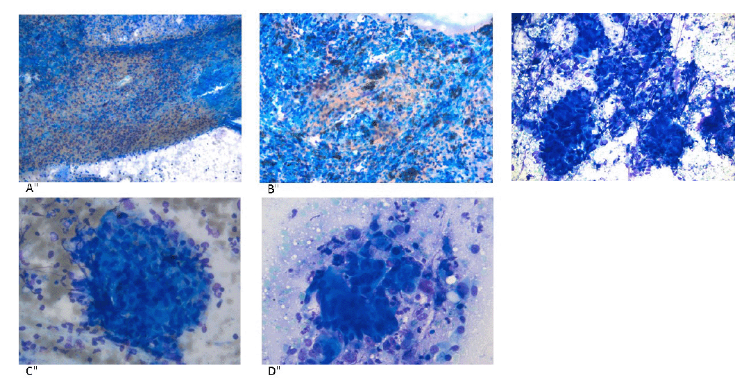
Slides are then stained by a cytotechnologist and reviewed by the cytopathologist on-site. Slides are considered adequate if evidence of tar-
get sampling was present. For example, when sampling a lymph node, the presence of lymphocytes, anthracosis, granulomas or tumor
would be considered adequate (Figure 1). Examples of inadequate samples include the presence of blood or benign bronchial cells (pick-ups)
only. If malignant cells are present, the tumor is then subtyped if possible based on cytomorphology alone. If the diagnosc subtype is fa-
vored to be non-small cell carcinoma, the smear is then evaluated to determine if adequate tumor is present for molecular studies. In our
molecular lab approximately 2000 tumor cells are required for Oncoscreen panel ( 50 genes) and 20,000 tumor cells for the OncoPlus panel
( > 1000 genes). The esmaon of cellularity is based on the experience of the cytopathologist, but more objecve tools should be applied
for clinical trials.
EBUS specimen handling for next generaon sequencing (NGS)
Jerey Mueller, MD
Associate Professor of Pathology
The University of Chicago
Tips from the Experts
P A G E 6 V O L U M E 5 , I S S U E 2
If more than half of the slide has tumor, the cellularity is considered adequate for both panels (Figure 2). If the smear is considered adequate
for diagnosis but inadequate for molecular studies, addional passes are performed and evaluated unl an adequate smear is obtained. If
needed, several smears may be combined to achieve the minimum cellularity requirement.
Molecular Pathology Evaluaon:
The selected smears are then submied to the molecular laboratory for next generaon sequencing (NGS). This tesng includes the lung
fusion panel (ALK/RET/ROS1 fusion gene tesng) and the Oncoscreen solid tumor mutaon panel, which includes all currently targetable mu-
taons in NSCLC
Conclusion:
In my opinion, using Rapid-Onsite Evaluaon in conjuncon with EBUS is far superior to a non-ROSE method for assessing adequacy for com-
prehensive molecular tesng and allows the most benet to the paent by minimizing excessive procedures, obtaining diagnosc and staging
informaon and adequate material for molecular tesng. This pracce adheres to the current lung cancer guidelines.
References
1. Enger DS et al. JNCCN. 2017; 15(4), 504–35
2. Shao D et al. Sci Rep. 2016; 6(1), 22338. hp://doi.org/10.1038/srep22338
3. Fischer AH et al. JCO. 2011; 29(24), 3331–2– author reply 3332–3. hp://doi.org/10.1200/JCO.2011.35.2534
4. Vincek V et al. Lab Invest. 2003; 83(10), 1427–35
5. Treece AL. et al. Cancer Cancer Cytopathol. 2016; 124(6), 406–14. hp://doi.org/10.1002/cncy.21699
Figure legends
A. Benign lymphoid cells
B. Benign lymphoid cells and anthracosis
C. Granuloma
D. Adenocarcinoma
E. Adequate tumor for molecular studies
E”
Humanitarian News
W A B I P N E W S L E T T E R P A G E 7
The World Bronchology Foundaon Connues to Help Physicians Save Lives and Expand Their Bronchoscopic
Pracce in Guayaquil, Ecuador
In 2012, the World Bronchology Foundaon (WBF) donated a exible bronchoscope to the Hospital de Especialidades - Teo-
doro Maldonado Carbo in Guayaquil, Ecuador. This Hospital serves most of the workers in Guayaquil, as well as many people
coming from afar to receive treatment. Despite numerous improvements in infrastructure and programs in previous years,
the Hospital did not have a bronchoscope. In addion to donang equipment, members of the WBF provided a week-long
bronchoscopy training program (conducted by Dr. Henri Colt & Dr. Silvia Quadrelli) in order to assist increasingly well-trained
Respiratory Medicine sta. Through these eorts, the WBF started a new era in Guayaquil, an era during which the Respira-
tory Medicine Unit was to provide bronchoscopic diagnoses to their many paents. Today more than 30 bronchoscopies are
performed by this unit every month.
Four years later and as part of a connuous growth of the public health care sector in Ecuador, the hospital obtained a new
videobronchoscope from the government. The WBF had been tracking clinical acvies related to the use of originally do-
nated equipment as well as the connuous educaon in bronchoscopy of many members of the Respiratory Medicine Unit.
As part of ongoing follow-up, Dr. Quadrelli (Vice-chair of the WABIP) spent three days in Guayaquil in December, 2016. The
purpose of this trip was to help physicians of the Respiratory Medicine Unit become familiar with new equipment and assist
in training sta in the performance of transbronchial biopsies, only occasionally performed before. Training consisted of dis-
cussing paent scenarios, reviewing paent-care plans, and helping doctors perform 10 procedures under supervision. As a
result, Drs. Ulloa, De Janon & Figueroa gained condence in their abilies to perform transbronchial biopsies, now incorpo-
rated into their daily bronchoscopic pracce.
The WBF is proud of its ability to follow-up with equipment donaon and companion training programs around the world.
The excellent use made of the originally donated exible bronchoscope to the Guayaquil group prompted a change in clinical
pracce, and expanded the abilies of Ecuadorean doctors to assist hundreds of paents with lung diseases. The Foundaon
congratulates members of the Respiratory Medicine Unit for the progress they make each year. Thanks to the eorts of
charitable donaons, educators, and physicians eager to improve paent care, The World Bronchology Foundaon connues
to be a unique channel through which respiratory care is improved for paents in many countries around the world.
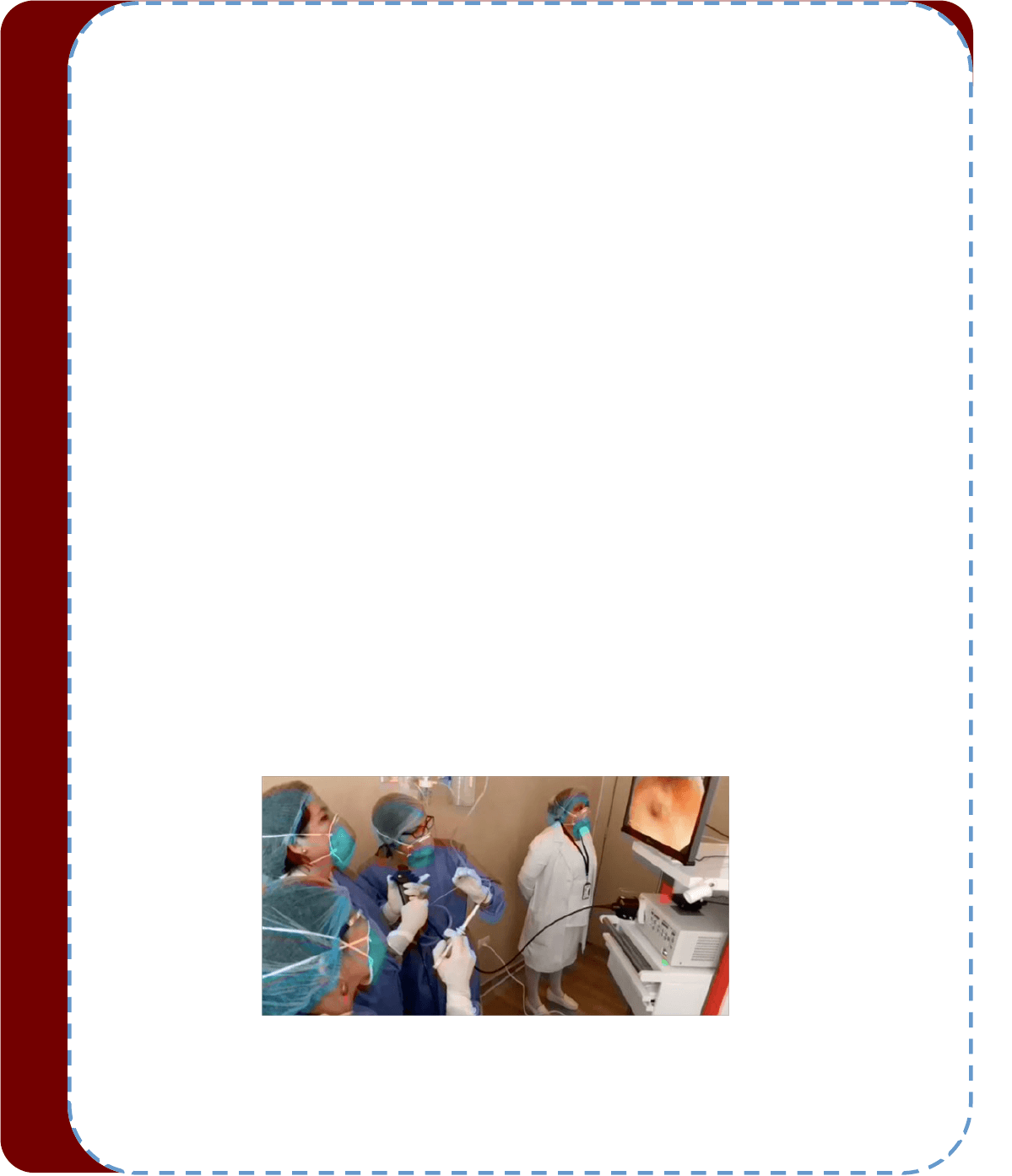
Figure 1: Physicians performing transbronchial lung biopsy using a new videobronchoscope in Guayaquil, Ecuador.
*The views expressed in this arcle are those of the author and do not necessarily reect the ocial posions of the Execuve
Board or Internaonal Board of Regents of the WABIP. Dr. Silvia Quadrelli is Vice-chair of the WABIP.
WABIP Train the Trainer and Introducon to Flexible Bronchoscopy Program, Maceio, Brazil 2017
Maceió, Brazil, a beauful city in the state of Alagoas, hosted the 2017 Train the Trainer and Introducon to Flexible Bronchoscopy program
conducted by the WABIP in April. Eleven leading bronchoscopists and university educators from several dierent regions in Brazil devoted ener-
gies to learning new educaonal methodologies and exploring their sense of movaon and dedicaon to changing the educaonal paradigm
in the country. As they became familiar with new teaching instruments such as the 4 box praccal approach, bronchoscopy assessment tools
and checklists, also employing role-playing exercises and applying step-by-step instruconal techniques, it became increasingly obvious that
change was needed. This was the second me an experience like this occurs in Brazil, allowing a new group of parcipants to get in touch with
Bronchoscopy Educaon Project philosophy. Priorizing “learner-centered” approach, models were used for “step-by-step” instruconal tech-
niques and to evaluate using competency-oriented validated assessment tools such as the BSTAT (Bronchoscopy Skills and Tasks Assessment
Tool). They had also the opportunity of using checklists to assure knowledge of me out, informed consent, and moderate sedaon, in conjunc-
on with simulaon scenarios and group exercises presented in the Bronchoscopy Educaon Training Manual.
Throughout this two and a half day seminar, didacc lectures, interacve sessions, group exercises, and key discussion points were facilitated
by Henri Colt (Immediate past Chair WABIP and author of The Essenal Bronchoscopy Series of books). A one-day Introducon to Flexible Bron-
choscopy program was held for sixteen Brazilian physicians in-pracce or in-training. This program provided opportunies for parcipants in the
Train the Trainer program to apply their newly learned skills and ideas. Increasing interacon between students and faculty is the key to ac-
quire cognive, aecve, and experienal knowledge.
This 2017 program was directed and organized by Master Instructor, Dr. Viviana Figueiredo (Sao Paolo), and Dr. Tadeu Lopez (Maceio). The
courses were ocially endorsed by the Brazilian Society for Thoracic Surgery (SBCT) and the Brazilian Society for Pneumology and Tisiology
(SBPT). Guest instructors included Hugo Oliveira (cered instructor, Porto Allegre, Brazil) and Patricia Vujacich (Chair of the WABIP Educaon
Commiee, Buenos Aires, Argenna).
Each Train the Trainers Seminar is dierent. This me, closing remarks on acve listening, learner centered approach, case based problem solv-
ing, role-playing educaonal techniques, presentaon skills, lecturing and condenal self-assessment made it exceponal in terms of mova-
on. Iniaves of translang into Portuguese several materials of Bronchoscopy Internaonal were undertaken. So far, numerous university
programs have adopted BSTAT, Step-by-Step, Praccal Approach, and the Informed Consent Checklist into their training programs.
As soon as more learning materials become available in Portuguese it is our hope to expand training to other regions in 2018, and to connue
to disseminate Bronchoscopy Educaon Project philosophy and learning tools throughout Brazil.
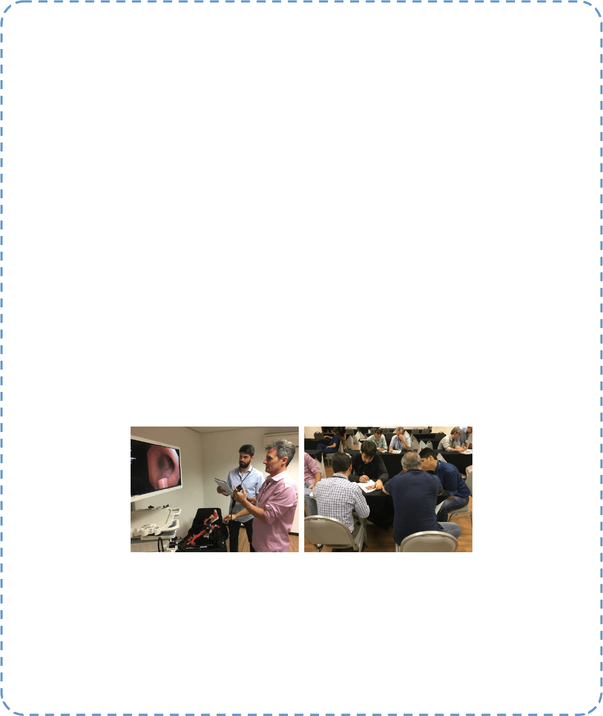
Figures 1 and 2 above: Learning to apply technical skill training Step-by-Step during the Train the Trainer program using BSTAT in an inanimate
airway model (Drs. and thoracic surgeons/bronchoscopists Filipe Andrade and Spencer Camargo). Small group workshops provide students in
the Introducon to Flexible Bronchoscopy course an opportunity to implement a common learning for secreon and mucosal ndings using
BSTAT.
Education and Training
P A G E 8
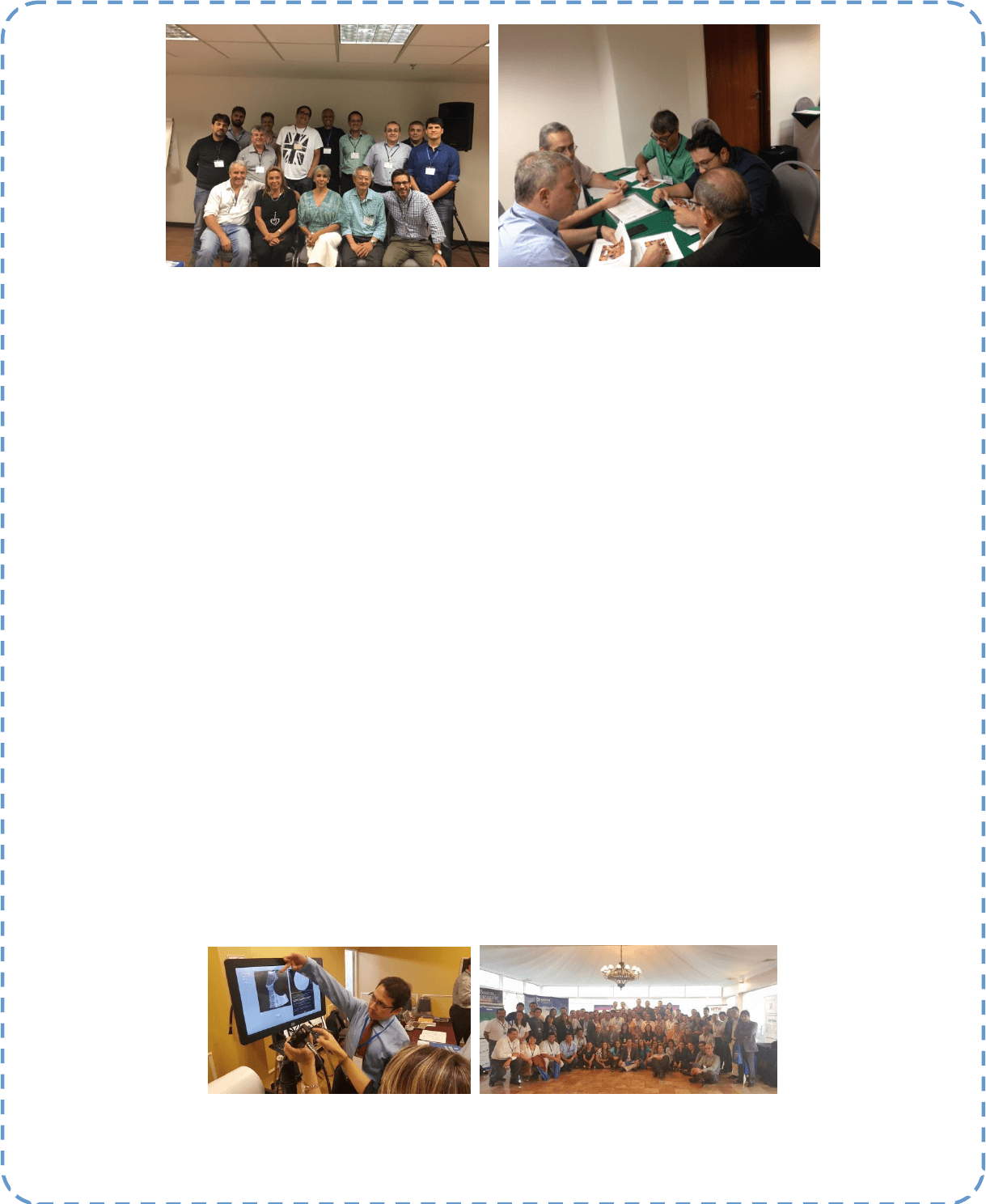
Figure 3: Opinion leaders from throughout Brazil gathered to parcipant in the 2017 Train the Trainers Program hosted by Dr. Vivian Figueiredo
(sing, middle) and Dr. Tadeu Lopez (standing right, dark blue shirt). Master Instructors Patricia Vujacich (Argenna) and Hugo Oliveira (Brazil)
are seated to the right of Vivian. Figure 4: Small group case-based praccal approach and BSTAT exercise during the Introducon to Flexible
Bronchoscopy program held in Maceio, Brazil.
XI Biennial Congress of the South American Society for Respiratory Endoscopy (ASER)
This year’s congress was held in Lima, Peru hosted by ASER President Hugo Boo (Argenna) and Congress President Pedro Garcia Manlla
(Peru). A record number of parcipants from throughout Lan America came to Lima for three days of collegiality, friendship, and scienc
engagement. The program included a sponsored Symposium on Thoracic Oncology, chaired by Dr. Silvia Quadrelli, Vice-Chair of the WABIP,
with parcipaon of known Oncologists (Brian Hunnis from Florida University and Carlos Silva from Argenna), a Peruvian intervenonal pul-
monologist on sta at Henry Ford Hospital (Dr. Javier Diaz-Mendoza) and a Peruvian Radiaon Oncologist (Gustavo Sarria).
With more than 100 aendees, congress parcipants included dozens of foreigners from Argenna, Bolivia, Brazil, Chile, Colombia, Spain, Para-
guay, and the United States, as well as 66 Peruvians (58 from Lima and 8 from the Provinces). Faculty numbered 35 speakers and instructors of
which 23 came from abroad and 12 were from Peru. Overall, 72 people aended hands-on workshops organized by Dr. Fernando Monge from
the Peruvian Associaon for Bronchology and Dr. Javier Diaz-Mendoza, with generous support from Industry. Conference aendees unani-
mously say they le Lima with renewed enthusiasm and knowledge of new approaches and techniques that improve their clinical pracce.
Workshop sessions included:
Dicult Airway and Pediatric Bronchoscopy
PDT, Percutaneous Cricothyrotomy, and Bronchoscopy Intubaon
Interacve Sessions: Informed Consent and Praccal Approach exercises for cTBNA
Mediasnal Anatomy and EBUS TBNA
Intervenonal Pleura Procedures and Thoracic Ultrasonography
Rigid Bronchoscopy Intubaon, Stent Placement
Central Airway Obstrucon: Electro and Cryosurgery, including Foreign Body Removal
During the ASER’s business meeng, Chile was chosen to host the next ASER XII Congress. Similar to regional bronchology associaon meengs
in Europe and Asia, the ASER meeng will now be held every two years interposed with the Biennial World Congress of the WABIP. Also during
the congress, the WABIP held a meeng for all South American regents. News from this meeng will be announced in the WABIP Newsleer,
including iniaves proposed by Regents to further the adopon of Bronchoscopy Educaon Project materials across Lan America, and ways
to enhance Regents’ parcipaon in both global and regional acvies of the WABIP.
Figure 1: Dr. Javier Diaz-Mendoza teaching at the EBUS workstaon. Figure 2: Parcipants and faculty at the hands-on workshops of ASER, Peru
2016.
Education and Training
P A G E 9

Board of Regents News
(Le to right: Dr. Bilaceroglu, Dr. Encheva, Dr. Flandes, Dr. Arshad Husain, Dr. Musani and Dr. Niwa)
WABIP NEWS
P A G E 10
NEW Board of Regents Members (updated April 2017) - The WABIP is honored and pleased to welcome six new members on the
Board of Regents. The new Regents are doctors: Semra Bilaceroglu (Turkey – EABIP), Milena Encheva (Bulgaria), Javier Flandes
(Spain – AEER), Syed Ar- shad Husain (UK – BBG), Ali Musani (USA – AABIP) and Hiroshi Niwa (Japan – JSRE)
WABIP Rare Lung, Airway and Pleura Disorders - A brand new Facebook group for this section is currently live. We invite you to
join this growing group, with now 127 participants from around the world. Click the below link to begin:
https://www.facebook.com/groups/WABIPrarelungairwaydisorders/
Featured WABIP Member Society - Founded in 2002 by Dr. Heinrich Becker, Dr. Chris Bolliger and Dr.
Felix Herth, the European Associaon for Bronchology and Intervenonal Pulmonology (EABIP) is
dedicated to the promoon of high standards in clinical pracce, educaon and research in diagnosc
and therapeuc intervenonal pulmonology including bronchoscopy and thoracoscopy, as well as in the
educaon and training of endoscopy sta, in Europe. With the ECBIP congress just wrapping up only
weeks ago to an astounding success, we invite you to learn more about the society, its leaders and
members. More at hp://www.eabip.org/
New WABIP website – We are pleased to present to you a brand new website funconalies & design that takes the WABIP for-
ward as one of the leading medical associaons in the digital and mobile era. Without further ado, have a look at the new site at
hp://www.WABIP.com
WABIP Pediatric Bronchoscopy – You are cordially invited to join our new Pediatric Bronchoscopy WhatsApp Group , which has
154 parcipants on board at the me of this wring. Click the following link on your WhatsApp enabled mobile device to join:
https://chat.whatsapp.com/K1BawQYGOfjDTF6ljfMg19
With over 50 members strong, the WABIP Board of Regents is the principal governing body of our organizaon. Members of the
BOR vote on Vice-chair candidates in biennial elecons, Bylaws amendments, WCBIP host candidates and other maers central to
governing the WABIP. The BOR is comprised of all members on the WABIP Execuve Board and a minimum of one representave
from every WABIP Member Society.
Annual Board of Regents Meeng –We are proud to announce that the annual meeng held in February 2017 was a success, and
that the business items of reviewing and vong on the prior year and current year acvity & nancial reports were carried out
eecvely. We wish to thank all 29 (out of 52) parcipang Regents in providing valuable eort and support in execung the mis-
sion of the WABIP.
Endobronchial Ultrasound Guided Transbronchial Needle Aspiraon: Beer than Transbronchial
Biopsy for PD-L1 Proling in Lung Cancer
Several recent studies have raised the queson of validity of Endobronchial Ultrasound Guided Transbronchial Needle Aspiraon (EBUS –TBNA)
in sampling for Programmed Death Ligand 1 (PD-L1). The quesons spanned from the amount of cells obtained to the severity of crushed cells
rendering specimen uninterpretable. It was suggested that perhaps the surgical specimens were beer for its size and minimal crushing of the
ssue.
Well, EBUS-TBNA has shown its dominance in ssue sampling yet again. In samples obtained from lymph nodes in the paents with lung mass-
es who also underwent Transbronchial Biopsy (TBBX) and in some cases resecon of the lesion, the number of cells obtained were signicantly
more than the TBBX and the amount of crushed cells were signicantly less than TBBX. Hence, oering a much beer quality specimen for PD-
L1 analysis. The specimens obtained by EBUS-TBNA also showed a high concordance rate with the surgical specimens in both, primary tumors
and metastac diseases (1).
A recent study from Japan (1), prospecvely looked at 97 paents with EBUS-TBNA specimens, 20 of whom also had TBBX done. These speci-
mens were evaluated for PD-L1 expression as well as the morphological health of the cells (lack of crush/destrucon eect). The study showed
that the total number of cells obtained from EBUS-TBNA were stascally signicantly (P<.001) more than the cells obtained from the TBBX
and the crush eect on biopsy samples was also signicantly lower (P<.001) than TBBX. The PD-L1 expression on EBUS-TBNA specimen showed
good concordance rate with the TBBX, primary tumor and the metastac nodes.
The ability of EBUS-TBNA in providing large number of high quality cells with minimal crush eect and its feasibility for broad spectrum molecu-
lar assays including EGFR (Epidermal Growth Factor Receptor), ALK (Anaplasc Lympho-Kinase) (2)(3) and the likes and now PD-L1 conrms it
as a robust, minimally invasive, high yield, cheap, and quick test for lung cancer diagnosis, staging, and personalized therapy. With the growing
numbers of aconable bio-markers such as above menoned, the signicance of EBUS-TBNA is limitless.
References
1. Sakakibara R et al. cllc.2016.12.002
2. Navani N et al. Am J Respir Crit Care Med 2012; 185:1316-22.
3. Nakajima T et al. Chest 2007; 132:597-602
Editorial Staff
Associate editor: Dr. Ali Musani
Associate editor: Dr. Sepmiu Murgu
Editor-in-Chief: Dr. Kazuhiro Yasufuku
Research
Primary Business Address:
Kazuhiro Yasufuku, Editor-in-Chief
WABIP Newsleer
c/o Judy McConnell
101 College St., TMDT 2-405
Toronto, Ontario M5G 1L7
Phone: 416-581-7486
E-mail: newsleer@wabip.com
P A G E 11
P A G E 12
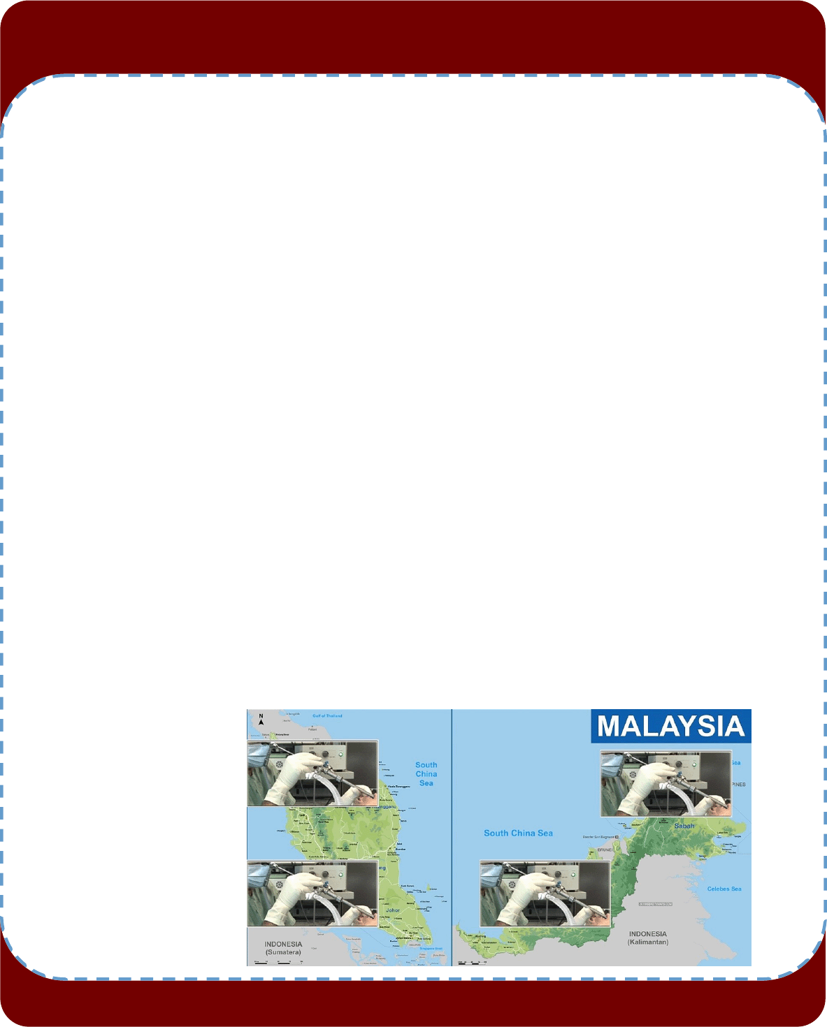
Malaysia has an esmated populaon of 31 million. There are about 60 pulmonologists in the country.
The Malaysian Associaon for Bronchology and Intervenonal Pulmonology (MABIP) was founded on 31
st
December 2013 and has a
membership of about 90 local and overseas members.
In 2007, a group of local pulmonologists organised the rst Naonal intervenonal bronchoscopy course. Before 2007, intervenonal
bronchoscopy in Malaysia was patchy and there was no concerted eort to promote the experse. Most pulmonologists then per-
formed basic diagnosc bronchoscopy. Cases that required intervenonal approach would be referred to thoracic surgeons. Encouraged
by the overwhelming response, the intervenonal course was held annually and always had excellent parcipaon from South East Asia
delegates. In 2013, it was felt that a society dedicated to intervenonal pulmonology was needed to promote the growth and pracce in
Malaysia. This annual event which started inially as a course in 2007 has now become a fully edged scienc meeng under the MA-
BIP.
Bronchoscopy is included in the curriculum of the local pulmonary medicine fellowship. Since 2015, the MABIP runs an annual assembly
for pulmonologists specically devoted to intervenonal pulmonology. It includes didacc lectures, symposia, hands-on workshops, live
cases and free papers. This annual assembly is also aended by pulmonologists from all over Asia. Training in rigid bronchoscopy is
oered by 4 centres, 2 in West Malaysia and 2 in East Malaysia (photo aached).
Basic bronchoscopy, pleuroscopy (including rigid thoracoscopy) and convenonal TBNA are performed in most university and public
hospitals in Malaysia. More than 90% of bronchoscopists are pumonologists. Some general physicians are given privileging in basic bron-
choscopy. As rigid bronchoscopy is only available in 4 centres, other hospitals usually refer their cases to these 4 centres. Therapeuc
bronchoscopy (debulking, airway dilataon, stent placement, electrocautery) is available in these 4 centres. Currently, only 1 centre
performs addional advanced diagnoscs and therapeucs (cryobiopsy, electromagnec navigaon, bronchial thermoplasty and endo-
bronchial valves). 9 centres have linear EBUS and 1 centre has a radial EBUS and a YAP laser respecvely.
The main indicaons for bronchoscopy in Malaysia are in the invesgaon of hemoptysis and lung cancer. Tuberculosis is endemic in
Malaysia and therefore it is sll an important diagnosc indicaon. Sedaon is rounely performed in all instuons, in some centres
using TIVA with the cooperaon of anaesthesiologists for rigid bronchoscopy.
The strong support from the WABIP has allowed the MABIP to conduct its annual assembly and promote the growth of intervenonal
pulmonology in Malaysia and Asia.
BRONCHOSCOPY AROUND THE WORLD
W A B I P N E W S L E T T E R
P A G E 12
P A G E 13
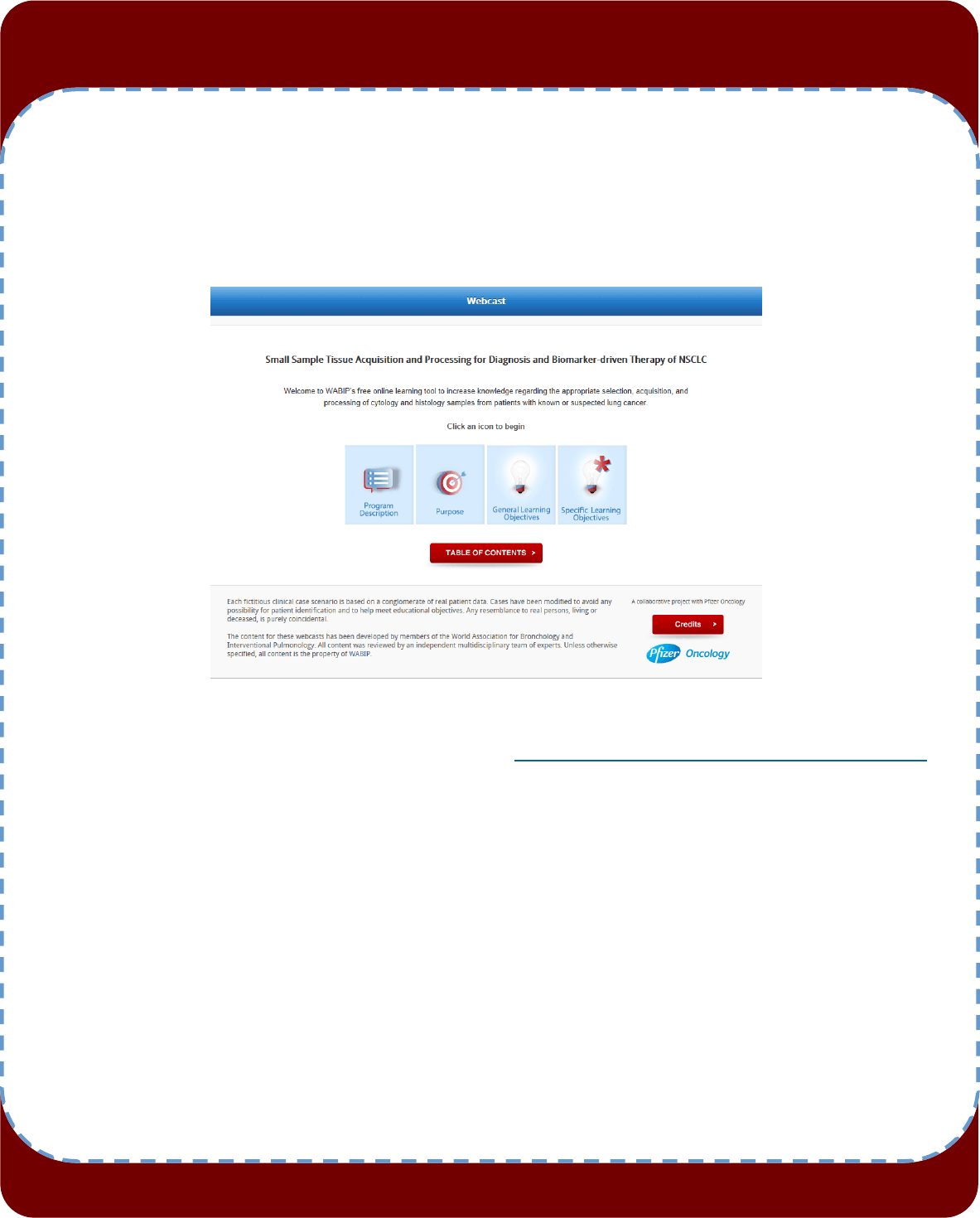
WABIP ACADEMY- WEBCASTS
The WABIP has started a new educaon project recently: THE WABIP ACADEMY. The WABIP Academy will pro-
vide free online webcasts with new and hot topics that will interest pulmonologists and intervenonalists.
Current webcast topic: Tissue acquision for biomarker directed therapy of NSCLC
You can reach these webcasts by using this link: hp://www.wabipacademy.com/webcast/
www.bronchology.com Home of the Journal of Bronchology
www.bronchoscopy.org Internaonal educaonal website for
bronchoscopy training with u-tube and
facebook interfaces, numerous teachiing
videos, and step by step tesng and assess
ment tools
www.aabronchology.org American Associaon for Bronchology and I
ntervenonal Pulmonology (AABIP)
www.eabip.org European Associaon for Bronchology and
Intervenonal Pulmonology
W A B I P N E W S L E T T E R
Links
www.chestnet.org Intervenonal Chest/Diagnosc Procedures (IC/DP)
NetWork
www.thoracic.org American Thoracic Society
www.ctsnet.org The leading online resource of educaonal and
scienc research informaon for cardiothoracic
surgeons.
www.jrs.or.jp The Japanese Respirology Society
sites.google.com/site/asendoscopiarespiratoria/
Asociación Sudamericana de Endoscopía Respiratoria
P A G E 13

P A G E 14
UPCOMING EVENTS
EBUS and Advanced Diagnosc Bronchoscopy: The Sixth Year (MD, USA)
When: July 20-21, 2017
Where: Hya Regency Chesapeake Bay, Cambridge, MD
Program Director: Lonny Yarmus, DO, MD
Program Type: Educaonal seminar (postgradua,te may include physicians in pracce and trainees)
Hands-on workshop, Conference (didacc lecture 3rd Annual MABIP Assembly (Malaysia))
When: 3-5 OCTOBER 2017
Where: LE MERIDIEN PUTRAJAYA, MALAYSIA
Program Director: ROSMADI ISMAIL, MD
Program Type: Hands-on workshop, Conference (didacc lectures)
Asian-Pacic Congress on Bronchology and Intervenonal Pulmonology 2017 (Indonesia)
When: November 2-4, 2017
Where: Ayodya Nusa Dua Bali, Indonesia
Program Director: Wahyu Aniwidyaningsih, MD, PhD, MD
Program Type: Educaonal seminar (postgraduate may include physicians in pracce and trainees)
Hands-on workshop Conference (didacc lectures)
W A B I P N E W S L E T T E R
P A G E 14
Introducon to Flexible Bronchoscopy (La Habana, Cuba)
3rd Annual MABIP Assembly (Malaysia)
Date: June 9-10, 2017
Venue: Hospital Neumológico de la Habana. Universidad de Medicina Salvador Allende. La Habana, CUBA
Program Directors: Manuel Sarduy, MD and Patricia Vujacich, MD
Program Type: Bronchoscopy course - Introducon to Flexible Bronchoscopy
Introduction to Flexible Bronchoscopy (Bolivia)
Date: June 1 - 2, 2017
Location: Sucre - Bolivia
Program Director: Edgar Benitez-Anibarro, MD
Program Type: Bronchoscopy course - Introduction to Flexible Bronchoscopy